Once upon a time, in the late 16th century, the world was filled with mysteries that lay beyond the reach of human vision. Scientists and scholars yearned to explore the hidden realms of the tiny, the unseen, and the unknown. It was during this era that the story of the compound microscope began.
Our tale begins with two Dutch spectacle makers, Hans Jansen and his son Zacharias, who are often credited with inventing the compound microscope around the year 1590. Legend has it that they accidentally discovered the power of lenses to magnify small objects while experimenting with their lenses in their workshop in Middelburg, Netherlands.
Word of this groundbreaking invention quickly spread throughout Europe, capturing the imaginations of scientists, scholars, and explorers alike. Soon, skilled craftsmen began refining and improving upon the design, leading to the development of more sophisticated and powerful microscopes.
One of the key figures in the advancement of the compound microscope was the Dutch scientist Antonie van Leeuwenhoek. In the late 17th century, using microscopes of his own design and construction, Leeuwenhoek became the first person to observe and describe microorganisms, including bacteria, protozoa, and sperm cells. His groundbreaking discoveries revolutionized our understanding of the natural world and laid the foundation for the field of microbiology.
As the centuries passed, the compound microscope continued to evolve, with advancements in optics, magnification, and illumination enhancing its capabilities. By the 19th century, the microscope had become an indispensable tool in scientific research, education, and medicine, allowing scientists to explore the intricacies of cells, tissues, and microorganisms in unprecedented detail.
In the modern era, compound microscopes are found in laboratories, classrooms, and research institutions around the world, enabling scientists to unlock the secrets of the microscopic world and push the boundaries of human knowledge. From unraveling the mysteries of disease to exploring the wonders of the natural world, the compound microscope remains a symbol of humanity’s insatiable curiosity and relentless pursuit of discovery. And so, the story of the compound microscope continues, illuminating the unseen and inspiring future generations to explore the vast wonders that lie beyond the limits of the naked eye.
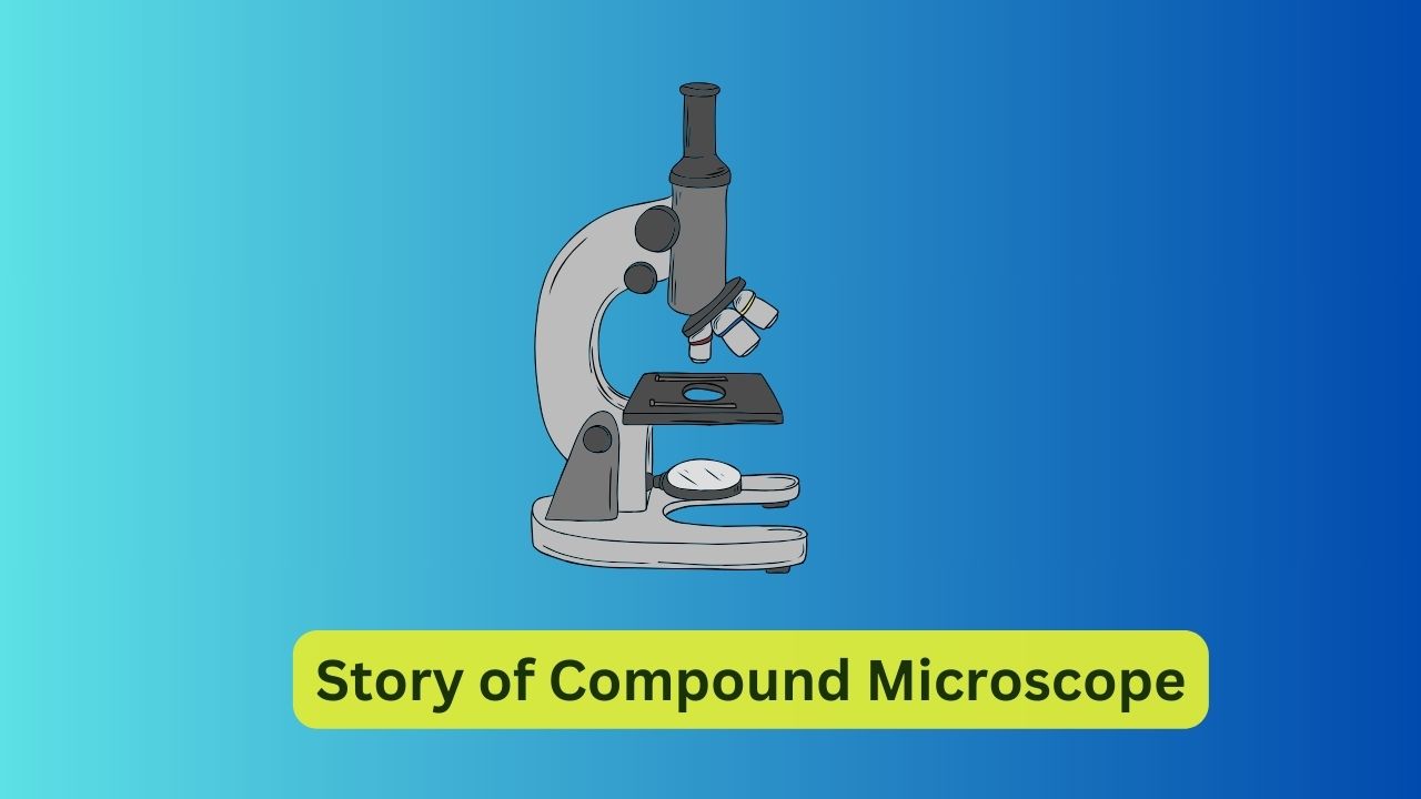
A compound microscope is an optical instrument used for viewing objects that are too small to be seen with the naked eye. Here are its main parts, functionalities, and usage:
Parts of a Compound Microscope:
- Base: The bottom part of the microscope that provides stability.
- Arm: The vertical support that connects the base to the head.
- Head: Also known as the body tube, it holds the optical components and eyepieces.
- Eyepiece: The lens closest to the eye, often magnifying the image by 10x.
- Objective Lenses: Lenses positioned close to the specimen, usually available in multiple magnifications (e.g., 4x, 10x, 40x, 100x).
- Stage: The platform where the specimen is placed for observation.
- Stage Clips: Clips to hold the specimen slide in place on the stage.
- Condenser: Concentrates light onto the specimen for better illumination.
- Iris Diaphragm: Controls the amount of light passing through the condenser.
- Coarse Adjustment Knob: Moves the stage up and down for rough focusing.
- Fine Adjustment Knob: Allows for precise focusing.
- Light Source: Typically located beneath the stage, provides illumination for the specimen.
- Mechanical Stage: Allows for precise movement of the specimen slide.
Functionalities:
- Magnification: Compound microscopes offer variable magnification through interchangeable objective lenses.
- Resolution: Provides high-resolution images, allowing for the visualization of fine details.
- Illumination: Illumination systems, such as a built-in light source and condenser, ensure proper lighting of the specimen.
- Focusing: Allows for both coarse and fine adjustments to bring the specimen into sharp focus.
- Observation: Used for observing various biological specimens, cells, tissues, microorganisms, and other minute structures.
- Analysis: Enables detailed analysis and study of microscopic structures.
- Photography and Recording: Some compound microscopes are equipped with attachments for photography and video recording of specimens.
- Research and Education: Widely used in scientific research, education, medical diagnosis, and industrial applications.
Usage:
- Prepare the Specimen: Place the specimen on a glass slide and cover it with a coverslip if necessary.
- Adjust Illumination: Adjust the intensity and focus of the light source to illuminate the specimen adequately.
- Select Objective Lens: Start with the lowest magnification objective lens (usually 4x) and gradually increase magnification as needed.
- Focus the Specimen: Use the coarse adjustment knob to bring the specimen into rough focus, then fine-tune using the fine adjustment knob for sharp focus.
- Observe and Analyze: Once the specimen is in focus, observe and analyze its details under the microscope.
- Capture Images or Record: If needed, capture images or record videos of the specimen for further analysis or documentation.
- Clean and Maintain: After use, clean the lenses and stage, and store the microscope properly to maintain its functionality.
Compound microscopes are indispensable tools in various scientific fields, providing invaluable insights into the microscopic world.
No responses yet